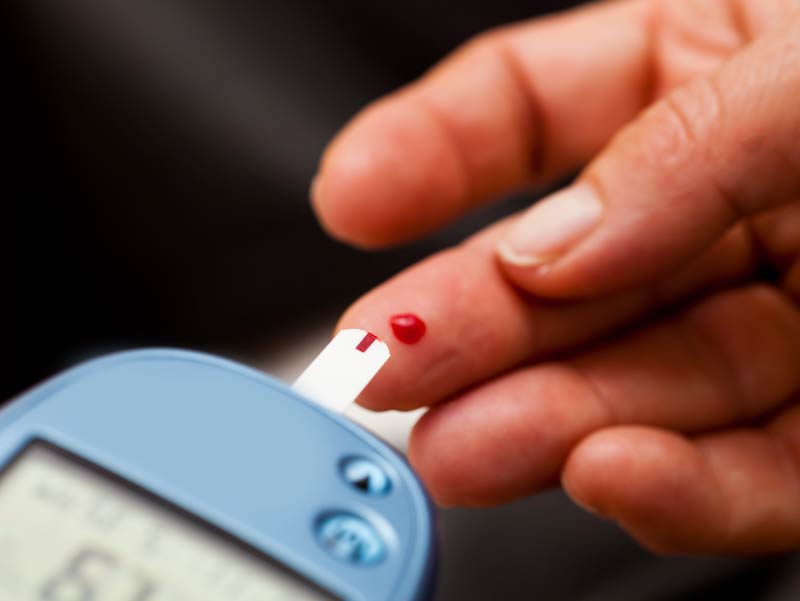
From its historical origins in ancient Egypt to modern-day diagnostic challenges, diabetes mellitus remains a complex disorder impacting global health. This condition, characterized by disturbances in glucose homeostasis, underscores the critical role of insulin and its intricate metabolic pathways.
BY DR. T . VISHNU MURTHY
Overview
Diabetes mellitus is a diagnostic term encompassing a range of anatomical and biochemical abnormalities that share a common disturbance in glucose homeostasis, primarily due to a deficiency in the beta cells of the endocrine pancreas. This definition, while broad and somewhat vague, reflects the significant variability of the disorder. Similar to syphilis, diabetes and its complications impact numerous areas of medicine.
The syndrome of diabetes mellitus can be asymptomatic, or it may present as a disorder affecting any organ or system. Fulminant ketoacidosis, which is fatal unless treated immediately, may be the initial sign. More often, diabetes manifests through long-term complications such as foot ulcers, retinopathy, or proteinuria. Additionally, other pathological states that are more frequent in diabetics compared to the general population may provide clues. For instance, a myocardial infarction in a young man, an unexpectedly large newborn, or recurrent skin infections can be indicative of diabetes. This condition is protean in its manifestations, making its diagnosis and treatment particularly challenging.
Carbohydrate Reserves and Fuel Utilization
In terms of energy storage, the human body has approximately 70 grams (280 kilocalories) of liver glycogen and 200 grams (800 kilocalories) of muscle glycogen. The glucose present in body fluids is even less, about 15 to 20 grams. With slightly more than 1 kilocalorie per minute required for basal metabolic needs in a resting adult, the total available carbohydrate in the body provides less than a day’s supply of fuel. Unlike carbohydrates and fats, proteins do not have a dedicated storage depot; they are all used for structural purposes, enzymes, or other essential functions. These points underscore the role of fat as the primary energy storage depot in humans and the minimal role of carbohydrates.
Tissue Fuel Utilization
Despite fat being the predominant storage form of energy, certain tissues, particularly the brain, require a constant supply of glucose. Therefore, glucose concentration in a normal individual is maintained between 50 and 150 mg per 100 ml—levels above the minimum required by the brain but below the saturation point of renal reabsorption capacity. Insulin plays a crucial role in maintaining glucose within this range. While all tissues can use glucose when it is abundant in the diet, they only do so if insulin levels are high. Tissues like fat and muscle do not utilize glucose when insulin levels are low, instead relying on free fatty acids released from adipose tissue. Hence, the endocrine pancreas’s capacity to respond quickly and adequately to nutritional states by regulating insulin release is critical for fuel homeostasis.
The Fed State
After consuming a mixed meal, the gastrointestinal tract digests proteins into amino acids and carbohydrates into simple sugars, which then enter the portal bloodstream. The liver, which is permeable to all simple sugars and related molecules, has specific enzymes for the phosphorylation and metabolism of these sugars, particularly galactose and fructose. Glucose is phosphorylated and either incorporated into glycogen or metabolized by the liver for its energy needs and fat synthesis. These processes necessitate high insulin levels. Following a large carbohydrate-containing meal, much of the glucose also passes to peripheral tissues, with increased insulin levels facilitating glucose transport into muscle and adipose cells.
The third dietary component, triglycerides, is partially hydrolyzed in the gut, absorbed by the mucosa, re-synthesized into triglycerides, and incorporated with several lipoproteins into chylomicrons. These chylomicrons are secreted into the lymphatic system, eventually entering the general circulation via the thoracic duct.
Fasted State
During the fasted state, as described previously, humans possess minimal reserves of carbohydrates which are quickly depleted, even during overnight fasting. Between meals, glycogenolysis maintains glucose levels initially, but prolonged caloric deprivation necessitates hepatic gluconeogenesis. This process is initiated by reduced beta cell release of insulin in response to falling glucose levels, resulting in decreased insulin levels. Consequently, this low insulin state triggers several metabolic responses: 1) net muscle proteolysis, 2) release of amino acids to the liver via circulation, and 3) gluconeogenesis from these amino acids. The glucose produced is primarily utilized by the brain, although other tissues like the renal medulla, red blood cells, peripheral nerves, and to a limited extent, skeletal muscle also utilize glucose.

Diabetes
In the context of the metabolic states described above, abnormalities in insulin secretion lead to disruptions in fuel homeostasis. Inadequate insulin secretion following meal ingestion results in excessive intestinal absorption of all fuels, leading to postprandial hyperglycemia, hyperaminoacidemia, and hyperlipemia. This can be easily assessed through a glucose tolerance test, where oral or intravenous glucose administration reveals prolonged high blood glucose levels due to insufficient insulin release. Severe insulin deficiency further exacerbates the situation, impairing glucose uptake by muscle and adipose cells, promoting hepatic gluconeogenesis, and inhibiting hepatic ketogenesis. Consequently, glucose remains elevated in circulation, surpassing renal threshold and causing glucosuria, dehydration, hypovolemia, and severe hyperglycemia, potentially progressing to hyperosmolar coma.
In cases of severe insulin deficiency, irrespective of glucose concentration, peripheral amino acids are released from muscle proteins, providing the liver with abundant glucogenic substrates. This leads to accelerated gluconeogenesis and ketogenesis, reminiscent of prolonged starvation. Peripheral keto acid utilization decreases, causing rapid accumulation of beta-hydroxybutyrate and acetoacetate, which can precipitate life-threatening ketoacidosis alongside marked hyperglycemia, dehydration, and volume depletion.
Endocrine Pancreas
The complex disturbances in carbohydrate, fat, protein, and fluid metabolism observed in diabetic ketoacidosis closely resemble those induced experimentally by acute pancreatectomy, chemical destruction of beta cells with agents like alloxan or streptozotocin, or administration of insulin antibodies. Total or subtotal destruction of beta cells results in varying degrees of diabetes. Post-mortem examination of diabetic pancreases often reveals complete absence of beta cells in juvenile-onset diabetes or minor reductions with nonspecific morphologic changes in maturity-onset diabetes.
Beta cells respond to ambient glucose concentrations by releasing insulin in two phases: an immediate release peaking within 5 to 10 minutes, followed by a sustained release lasting 15 to 30 minutes as long as glucose levels remain elevated.
Insulin
Insulin is synthesized within beta cells as a single-chain proinsulin consisting of 86 amino acids. Post-synthesis, proinsulin folds back on itself, forming sulfur bridges and creating insulin and a connecting peptide. These components are packaged into granules within the beta cell. Upon stimulation, these granules migrate to the cell surface where insulin and the connecting peptide are released into adjacent capillaries. Normally, a small amount of proinsulin is also released, although its activity is significantly lower than insulin itself. In conditions such as islet cell tumors or diabetes, the ratio of proinsulin to insulin may increase, aiding in diagnostic assessment.
In the bloodstream, insulin circulates unbound, with approximately half being metabolized by the liver upon passage and the remainder eliminated via renal filtration and tubular catabolism. Its short half-life of approximately ten minutes suggests that changes in insulin concentration primarily reflect alterations in beta cell secretion rather than removal rates.
History
The description of diabetes dates back over 3000 years to ancient Egypt. Around 400 B.C., Charak and Susrut in India noted the sweetness of urine and observed correlations between obesity and diabetes, as well as its familial inheritance. They distinguished between two types of the disease: one associated with emaciation, dehydration, polyuria, and lethargy, and another characterized by its sweetness. In the Christian Era, Romans such as Aretaeus and Celsus described the disease, coining the term diabetes (meaning “siphon”) mellitus (from Latin “mellītus,” meaning “honey” or “sweet”).
The views expressed in this article are solely those of the author and do not necessarily reflect the opinions or views of this Magazine. The author can be reached at 9650696341

Leave a Reply